

Breast Diseases
Breast Tissue
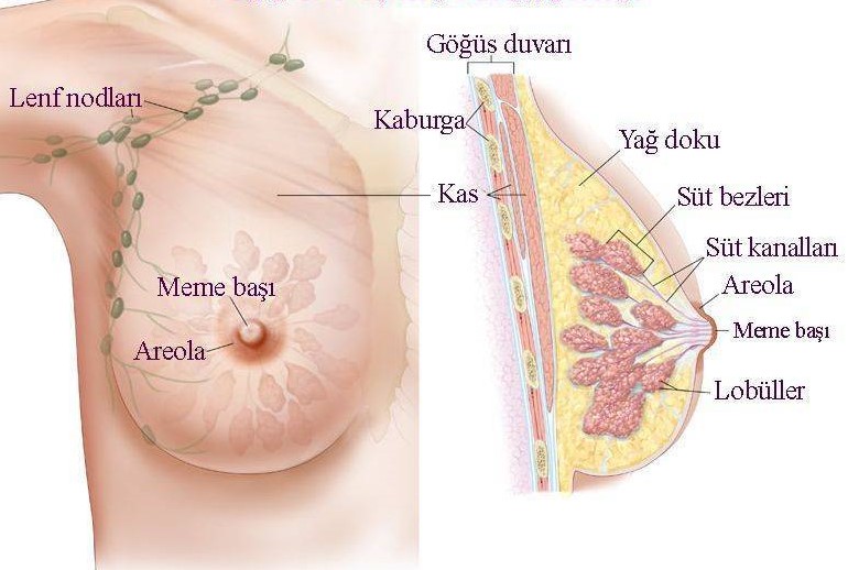
Approximately 15-20% of the breast tissue consists of glands that secrete milk in lactation, which we call the glandular structure.
1. General Information
The breast has been evaluated as a symbol of femininity beyond being just an organ. Its importance is undeniable in terms of aesthetics as well as in women's perception of bodily integrity. On the other hand, it also has a special importance in women, which creates an emotional-social bond between mother and baby. In men, on the other hand, the issue of whether it is purely healthy, as it remains a rudimentary, comes to the fore. Whatever way it is evaluated, it is bad. benign or benign diseases can cause serious problems.
Breast shape and size It is closely related to genetic, racial, dietary factors, number of pregnancies and menopausal status. Breast tissue can be cleft-hemispheric, conical, pendulum, thin or flat in varying anatomical shapes. It starts from the 2nd or 3rd rib on the chest wall and exhibits a unity with the component we call the nipple (nipple areolar complex) along the mid-axillary line extending to the 6th rib. It also extends to provide continuity with the armpit tissue on the outer parts of the body. This is also called the tail of the breast (tail of spence).
In the deep plane, the breast tissue lies on the M. Pectoralis Major fascia. It is also adjacent to the M. Serratus Anterior muscle, which is very important for respiration, and the M. Obliquus Externus muscle and the upper part of the M. Rectus Abdominis muscle sheath. As can be understood from these mixed definitions, in the vertical plane, the breast organ is above the chest wall, and the axillary area from the outside to the inside is quite wide between the collarbone and the abdominal wall. covers an area. Relatively loose lymph node between the breast tissue and many muscle and connective tissues on which it is located; and self-contained lymph nodes forms its own form with a connective tissue. These areas are called submammary-mammary tissue subspaces. Thanks to this structural feature, it is a mobile organ. This situation gains importance in the clinical staging of cancer, especially in deep-seated cancers, because of the decrease in breast mobility.
The nipple, which has an important function in the lactation period, which we call breastfeeding, is the "projection" in the middle of the breast and in the front part of the breast. It is a unit that completes the visuality we call. Depending on hormonal, developmental and neuroendocrine (pregnancy) factors, it can be in conical or flat shapes. It is located between the 4th rib in an anatomically healthy breast on the chest wall. The color of the nipple is closely related to the melanin pigment of the skin. In addition, this number of pregnancies varies according to racial characteristics.
2. Breast Skin
The breast organ is thin and unique. It is covered with a layer of skin and is covered with very thin hair follicles. However, the nipple has a special skin structure and has an indented, protruding, pigmented character. This region contains a large number of sweat and oil glands and is opened directly to the skin. The sebaceous glands on the nipple are softener-“lubricant” for the protection of the nipple during the breastfeeding period, which we call lactation. download it to make an impact. The tissues located between the sweat and the gland on the nipple grow during pregnancy and breastfeeding. "Melanocyte” especially from the 2nd month of pregnancy; The color begins to darken thanks to the cells called
a. Vascular nutrition and lymph drainage
Medially, the mammary skin is supplied by branches of the anterior intercostal arteries. These vessels then extend laterally to supply the intercostal muscles. Laterally, the mammary skin is supplied by the lateral thoracic artery, branches of the axillary artery, and lateral branches of the posterior intercostal arteries. The final drainage of the breast tissue, which is also provided with venous drainage by the veins accompanying the arteries, is to the axillary, internal thoracic and intercostal veins.
The density of lymph vessels belonging to the breast skin is higher than the other sides of the breast. Lymph drainage is predominantly to the armpit. The outer parts of the breast skin and the lymph drainage of the nipple are directed towards the pectoral lymph nodes. The interior of the breast skin The lymph drainage belonging to the parts of the body goes to the lymph nodes in the intercostal region. Lymph nodes in the parasternal area are interrelated in front of and behind the sternum, which should be kept in mind when planning the biological behavior and treatment of breast tumors.
3.Soft texture
The mammary tissue glands and small ducts containing the lobules that we call lobes can also be found in the channels.
It consists of tissue structure, which forms larger structures that take in. In the medical language, these are ordered from smallest to largest, as acini-lobule-TDLU-duct and lobule, and exhibit an architecture such that they are distributed over the quadrants in the breast. In direct proportion to the surgeon's experience, the lobe structure in the quadrants can be defined during the operation. This anatomical information is vital in determining the healthy surgical margin in breast-conserving surgery and determining the place where the appropriate tissue should be removed. “Lobule and lobe” Between these is the connective tissue formed by the collagen tissue (Figure 1). This connective tissue structure, which we call the stroma, is associated with the skin from the deep to the surface, from the lobules to the canals on the nipple. These connective tissue structures are more prominent especially in the upper quadrants of the breast and support the tissue. If this situation is involved by a tumor that may develop in the breast tissue, it is the cause of the symptom of pitting on the nipple or breast skin. There is adipose tissue between these breast lobules and lobes, which is important in the growth and shape of the breast under the influence of the hormone strogen in the developmental period we call puberty.
4. Vascular and lymphatic structure that provides nutrition to the breast tissue
Artery-Artery structures and Veins-Vein structures
Breast tissue is supplied by the axillary artery, internal thoracic artery and intercostal arteries. “venous plexus” around the nipple; There is a circular vein mesh that we call. Other vein structures accompany the arteries. These vascular structures are accompanied by the internal thoracic artery at the level of the 3rd intercostal cartilage tissue and "brachiocephalic" at the level of the clavicle. empties into the vein. This architectural structure should always be kept in mind by the surgeon in the biological behavior of breast tumors.
There are 2 lymphatic networks, superficial and deep, which are important in terms of giving an idea about the place where the cancer develops in the breast and its spread. Axillary lymph nodes collect 75% of the lymph drainage of the entire breast tissue. Between 20 and 40 lymph nodes in this region; has. It is divided into 3 groups as subscapular-central and apical. The lymph nodes on the pectoralis minor muscle are called Level 1, while those immediately adjacent to the muscle are Level 2, the lymph nodes located in the area between the upper edge of the muscle and the collarbone. Others are classified as Level 3 (Figure 2). Inside the breast The lymph nodes in the quadrants and some in the outer quadrants empty into the parasternal lymph nodes. These lymph nodes also show an upward course by making a network of interrelated lymph vessels on both sides of the breastbone. But it should not be forgotten that all lymphatics of the breast are always interrelated with a fine vascular network.
3. Surgical intervention for breast cancer and axillary lymph nodes
Surgery to the underarm lymph nodes is a part of breast surgery to be performed for cancer. &Cedil;∵ Which of the axillary regions we have listed above and classified as Level 1-2-3 has metastasis, how long is it? includes the lymph node, whether the tumor that metastasizes here goes beyond the lymph node, which part of the lymph node is involved will affect the chemotherapy and radiotherapy plans after surgery. Axillary lymphatic intervention, which is a part of breast surgery performed under the armpit and performed for cancer, can also cause undesirable effects such as loss of sensation-arm demi-arm movements-limitation of arm movements-post-operative fluid accumulation, which we call lymphocele.
4.Sentinel lymph node sampling
Today, to reduce the negative side effects of axillary lymph node dissection, to determine the minimum number of lymph nodes required for adequate staging, to save the patient from unnecessary underarm interventions It is a method used for Using a radioisotope material, a special dye is injected into the breast tissue from various anatomical regions before the surgery, using a special dye to create the "ink". lymph node appointment is made.